
קובץ:Electron micrograph of neuromuscular junction (cross-section).jpg
אין גרסה ברזולוציה גבוהה יותר.
Electron_micrograph_of_neuromuscular_junction_(cross-section).jpg (433 × 289 פיקסלים, גודל הקובץ: 95 ק"ב, סוג MIME: image/jpeg)
| זהו קובץ שמקורו במיזם ויקישיתוף. תיאורו בדף תיאור הקובץ המקורי (בעברית) מוצג למטה. |
.jpg)
.jpg?uselang=he)
תקציר
| תיאור |
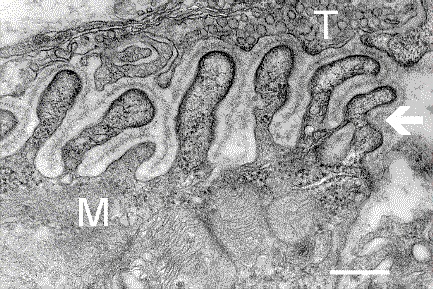
English: Electron micrograph showing a cross-section through the neuromuscular junction. T is the axon terminal, M is the muscle fiber. The arrow shows junctional folds with basal lamina. Postsynaptic densities are visible on the tips between the folds. The scale is 0.3 µm. |
| תאריך יצירה | Originally uploaded to en.wikipedia on 10 במרץ 2006. |
| מקור | Synapse Web at the National Institute of Mental Health, National Institutes of Health; originally from en.wikipedia; description page is/was here. |
| יוצר | National Institute of Mental Health; originally uploaded by Nrets at en.wikipedia. |
רישיון
This image is a work of the National Institutes of Health, part of the United States Department of Health and Human Services, taken or made as part of an employee's official duties. As a work of the U.S. federal government, the image is in the public domain.
 |
||
| הקובץ הזה זוהה כקובץ חופשי מכל מגבלה ידועה תחת חוק זכויות היוצרים, כולל כל הזכויות הקשורות והסמוכות. | ||
יומן העלאה מקורי
(All user names refer to en.wikipedia)
- 2006-03-10 20:07 Nrets 433×289×8 (97758 bytes) Electron micrograph showing a cross section through the neuromuscular junction. T is the axon terminal, M is the muscle fiber. The arrow shows junctional folds with basal lamina. Postsynaptic densities are visible on the tips between the folds. Scale is 0
היסטוריית הקובץ
ניתן ללחוץ על תאריך/שעה כדי לראות את הקובץ כפי שנראה באותו זמן.
| תאריך/שעה | תמונה ממוזערת | ממדים | משתמש | הערה | |
|---|---|---|---|---|---|
| נוכחית | 06:41, 22 במרץ 2007 | | 289 × 433 (95 ק"ב) | Fran Rogers | {{Information |Description=Electron micrograph showing a cross section through the neuromuscular junction. T is the axon terminal, M is the muscle fiber. The arrow shows junctional folds with basal lamina. Postsynaptic densities are visible on the tips be |
שימוש בקובץ
הדף הבא משתמש בקובץ הזה:
שימוש גלובלי בקובץ
אתרי הוויקי השונים הבאים משתמשים בקובץ זה:
- שימוש באתר ar.wikipedia.org
- שימוש באתר cs.wikipedia.org
- שימוש באתר de.wikipedia.org
- שימוש באתר en.wikipedia.org
- שימוש באתר es.wikipedia.org
- שימוש באתר fa.wikipedia.org
- שימוש באתר gl.wikipedia.org
- שימוש באתר ko.wikipedia.org
- שימוש באתר pt.wikipedia.org
- שימוש באתר ru.wikipedia.org
- שימוש באתר uk.wikipedia.org
- שימוש באתר zh.wikipedia.org
.jpg)