
קובץ:SEM blood cells.jpg

גודל התצוגה המקדימה הזאת: 482 × 600 פיקסלים. רזולוציות אחרות: 193 × 240 פיקסלים | 386 × 480 פיקסלים | 617 × 768 פיקסלים | 823 × 1,024 פיקסלים | 1,800 × 2,239 פיקסלים.
לקובץ המקורי (1,800 × 2,239 פיקסלים, גודל הקובץ: 1.33 מ"ב, סוג MIME: image/jpeg)
| זהו קובץ שמקורו במיזם ויקישיתוף. תיאורו בדף תיאור הקובץ המקורי (בעברית) מוצג למטה. |
תקציר
| תיאור |
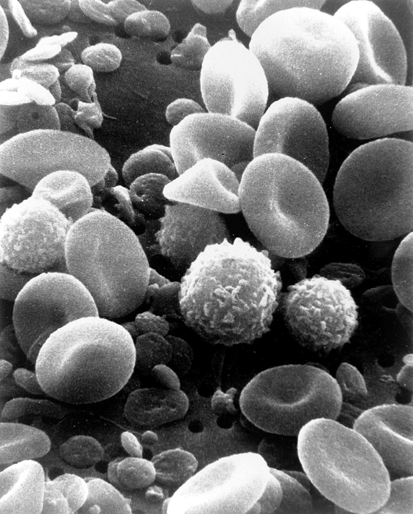
English: This is a scanning electron microscope image from normal circulating human blood. One can see red blood cells, several white blood cells including lymphocytes, a monocyte, a neutrophil, and many small disc-shaped platelets. Red cells are nonnucleated and contain hemoglobin, an important protein that contains iron and allows the cell to carry oxygen to other parts of the body. They also carry carbon dioxide away from peripheral tissue to the lungs where it can be exhaled. The infection-fighting white blood cells are classified in two main groups: granular and agranular. All blood cells are formed in the bone marrow. There are two types of agranulocytes: lymphocytes, which fight disease by producing antibodies and thus destroying foreign material, and monocytes. Platelets are tiny cells formed in bone marrow and are necessary for blood clotting. Type: Black & White Print Русский: Это изображение нормально циркулирующей крови человека получено с помощью сканирующего электронного микроскопа. Можно видеть красные кровяные тельца, несколько белых клеток крови (в их числе лимфоциты, моноциты, нейтрофилы) и множество мелких дискообразных пластинок. Красные кровяные тельца содержат гемоглобин — важный белок, который содержит железо и позволяет клетке переносить кислород к другим частям тела. Также они переносят углекислый газ от периферических тканей в лёгкие, где тот после газообмена может быть выдохнут. Лейкоциты борются с инфекциями и на две основные группы: гранулярные и агранулярные. Все клетки крови образуются в костном мозге. Есть два типа агранулоцитов: лимфоциты, которые борются с болезнью, производя антитела и тем самым разрушая чужеродный материал, и моноциты. Тромбоциты представляют собой крошечные клетки, образующиеся в костном мозге, и необходимы для свертывания крови. Тип фото: чёрно-белая печать. العربية : صورة بالمجهر الإلكتروني الماسح لدم الإنسان. يمكن للمرء أن يرى خلايا الدم الحمراء والعديد من خلايا الدم البيضاء بما في ذلك الخلايا الليمفاوية ووحيدات النوى والخلية المتعادلة والعديد من الصفائح الدموية الصغيرة ذات الشكل القرصي. |
||||||
| תאריך יצירה | Date Created: February 1982 | ||||||
| מקור | Image and description: National Cancer Institute | ||||||
| יוצר | Bruce Wetzel (photographer). Harry Schaefer (photographer) | ||||||
| אישורים והיתרים (שימוש חוזר בקובץ זה) |
|
||||||
| גרסאות אחרות |
Derivative works of this file: |
||||||

| Annotations | This image is annotated: View the annotations at Commons |
היסטוריית הקובץ
ניתן ללחוץ על תאריך/שעה כדי לראות את הקובץ כפי שנראה באותו זמן.
| תאריך/שעה | תמונה ממוזערת | ממדים | משתמש | הערה | |
|---|---|---|---|---|---|
| נוכחית | 21:17, 3 בפברואר 2021 | | 2,239 × 1,800 (1.33 מ"ב) | Tm | Reverted to version as of 20:27, 7 October 2006 (UTC) |
| 07:50, 10 בנובמבר 2020 |  | 2,239 × 1,800 (309 ק"ב) | Ratmanz | Optimized. | |
| 23:27, 7 באוקטובר 2006 |  | 2,239 × 1,800 (1.33 מ"ב) | DO11.10 | ||
| 06:00, 4 באוקטובר 2006 |  | 2,239 × 1,800 (989 ק"ב) | DO11.10 | {{Information |Description=This is a scanning electron microscope image from normal circulating human blood. One can see red blood cells, several white blood cells including lymphocytes, a monocyte, a neutrophil, and many small disc-shaped platelets. Red | |
| 04:09, 4 באוקטובר 2006 |  | 326 × 500 (36 ק"ב) | DO11.10 | {{Information |Description= A three-dimensional ultrastructural image analysis of a T-lymphocyte (right), a platelet (center) and a red blood cell (left), using a Hitachi S-570 scanning electron microscope (SEM) equipped with a GW Backscatter Detector. |
שימוש בקובץ
הדפים הבאים משתמשים בקובץ הזה:
שימוש גלובלי בקובץ
אתרי הוויקי השונים הבאים משתמשים בקובץ זה:
- שימוש באתר ar.wikipedia.org
- שימוש באתר ar.wikiversity.org
- שימוש באתר ast.wikipedia.org
- שימוש באתר as.wikipedia.org
- שימוש באתר az.wikipedia.org
- שימוש באתר ba.wikipedia.org
- שימוש באתר be-tarask.wikipedia.org
- שימוש באתר be.wikipedia.org
- שימוש באתר bg.wikipedia.org
- שימוש באתר bn.wikipedia.org
- שימוש באתר bn.wikibooks.org
- שימוש באתר bs.wikipedia.org
- שימוש באתר ca.wikipedia.org
- שימוש באתר ce.wikipedia.org
- שימוש באתר ckb.wikipedia.org
- שימוש באתר cs.wikipedia.org
- שימוש באתר cv.wikipedia.org
- שימוש באתר cy.wikipedia.org
- שימוש באתר de.wikipedia.org
- שימוש באתר de.wikibooks.org
- שימוש באתר dv.wikipedia.org
- שימוש באתר el.wikipedia.org
- שימוש באתר el.wiktionary.org
- שימוש באתר en.wikipedia.org