
קובץ:Human brain anterior-inferior view description.JPG
Human_brain_anterior-inferior_view_description.JPG (330 × 475 פיקסלים, גודל הקובץ: 31 ק"ב, סוג MIME: image/jpeg)
| זהו קובץ שמקורו במיזם ויקישיתוף. תיאורו בדף תיאור הקובץ המקורי (בעברית) מוצג למטה. |
תקציר
| תיאור |
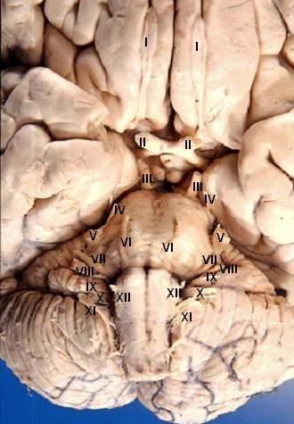
Human brain - anterior-inferior view - cerebral nerves I. N. olfactorius - The fila olfactoria (approximately 20 nerve rootlets on each side) comprise the Olfactory nerves (I) which run from the olfactory receptors in the nasal cavity to the olfactory bulb. II. N. opticus - The Optic nerves (II) are large & join at the midline to form the optic chiasm, then continue laterally as the optic tracts. The optic nerve is not a true nerve but rather a CNS tract. III. N. oculomotorius - The Oculomotor nerves (III) emerge from a depression in the midbrain, the interpeduncular fossa, just caudal to the optic chiasm. IV. N. trochlearis - The Trochlear nerves (IV) are small & are seen on the lateral surface of the midbrain. They are the only nerves which exit from the posterior side of the brain. V. N. trigeminus - The Trigeminal nerves (V) are large and emanate from the lateral surface of the pons. VI. N. abducens - The Abducens nerves (VI) exit near the midline from the inferior pontine sulcus which separates the pons from the medulla. VII. N. facialis - Moving laterally in the inferior pontine sulcus, the Facial nerves (VII) can be seen. VIII. N. vestibulocochlearis - Slightly lateral to the facial nerve is the Vestibulocochlear nerve (VIII). IX. N. glossopharyngeus - The postolivary sulcus is a groove running rostrocaudally on the lateral surface of the medulla. From this sulcus pass the small Glossopharyngeal nerves (IX) rostrally and... X. N. vagus - ...the much larger Vagus nerves (X) caudally. XI. N. accessorius - The Spinal Accessory nerves (XI) exit the cervical cord then pass rostrally through the foramen magnum to exit the cranial vault with the lossopharyngeal and vagus nerves. XII. N. hypoglossus - The Hypoglossal nerves (XII) exit the medulla via the preolivary sulcus.
|
| תאריך יצירה | |
| מקור | http://www.healcentral.org/healapp/showMetadata?metadataId=40566 (Internet Archive of file description page) |
| יוצר |
John A Beal, PhD Dep't. of Cellular Biology & Anatomy, Louisiana State University Health Sciences Center Shreveport |
| אישורים והיתרים (שימוש חוזר בקובץ זה) |
CC-BY |
| גרסאות אחרות |
 |
רישיון
- הנכם רשאים:
- לשתף – להעתיק, להפיץ ולהעביר את העבודה
- לערבב בין עבודות – להתאים את העבודה
- תחת התנאים הבאים:
- ייחוס – יש לתת ייחוס הולם, לתת קישור לרישיון, ולציין אם נעשו שינויים. אפשר לעשות את זה בכל צורה סבירה, אבל לא בשום צורה שמשתמע ממנה שמעניק הרישיון תומך בך או בשימוש שלך.
This file, which was originally posted to
http://www.healcentral.org/healapp/showMetadata?metadataId=40566, was reviewed on 25 September 2013 by reviewer Eleassar, who confirmed that it was available there under the stated license on that date.
|
היסטוריית הקובץ
ניתן ללחוץ על תאריך/שעה כדי לראות את הקובץ כפי שנראה באותו זמן.
| תאריך/שעה | תמונה ממוזערת | ממדים | משתמש | הערה | |
|---|---|---|---|---|---|
| נוכחית | 21:22, 24 ביוני 2006 | | 475 × 330 (31 ק"ב) | Patho | {{Information| |Description='''Human brain - anterior-inferior view - cerebral nerves''' I. N. olfactorius - The fila olfactoria (approximately 20 nerve rootlets on each side) comprise the Olfactory nerves (I) which run from the olfactory receptors in |
שימוש בקובץ
הדפים הבאים משתמשים בקובץ הזה:
שימוש גלובלי בקובץ
אתרי הוויקי השונים הבאים משתמשים בקובץ זה:
- שימוש באתר ar.wikipedia.org
- שימוש באתר azb.wikipedia.org
- שימוש באתר az.wikipedia.org
- שימוש באתר bs.wikipedia.org
- שימוש באתר de.wikipedia.org
- שימוש באתר de.wikibooks.org
- שימוש באתר en.wikipedia.org
- Cranial nerves
- User talk:Hovea
- User talk:Wouterstomp
- User talk:Nephron
- User talk:NCurse
- User talk:Robotsintrouble
- User talk:Was a bee
- User talk:Bloomingdedalus
- User talk:Read-write-services
- User talk:Attys
- User talk:Bakerstmd
- User talk:Manfi
- User talk:Mikepascoe
- User talk:Vokesk
- User talk:Neuraxıs
- User talk:Cmungall
- User talk:Qxukhgiels
- User talk:DocElisa
- User talk:JakobSteenberg/Archives/1
- User talk:Meteor sandwich yum
- User talk:Slashedone
- User talk:Anindya07
- User talk:Mattimussi
- User talk:Jelly Bean MD
- User talk:Benrusholme
- User talk:Brad.w.english
- User talk:Krsna ss
- User talk:Tom (LT)/Archive 4
- User talk:Dregon131
- User talk:Sapientia42
- User talk:Tilifa Ocaufa
- User talk:Snow Rise/Archive 6
- User talk:Arcadian
- User talk:Outofbattery
- User talk:Anatomyczar
- Wikipedia:WikiProject Anatomy/Newsletter/4
- User talk:Supravibhatsupravi
- User talk:Athikhun.suw
- User talk:CFCF/Archive 6
- User talk:Lewisskinner/ArchiveJul 2015
- User talk:Tyrol5/Archive 6
- User talk:CFCF/Archive 7
- User talk:DiverDave/Archive 5